Sudden increases in spring exercise are a leading trigger for cranial cruciate ligament tears in dogs. Learn to recognize the emergency signs, understand treatment options, and plan a safe rehabilitation timeline.
Key Takeaways
- Cranial cruciate ligament (CCL) tears often happen when sedentary dogs suddenly resume intense spring activities such as fetch, trail running, or off-leash play.
- A dog that is suddenly non-weight-bearing on a hind leg, especially after a burst of activity, should be treated as an urgent veterinary case.
- Immediate first aid involves strict rest, confinement, and safe transport: never attempt to splint, manipulate, or "pop" the joint back.
- Surgical repair (such as TPLO or lateral suture) is considered the gold standard for most dogs, though conservative management may suit select cases.
- Full rehabilitation typically spans 12 to 16 weeks and requires structured physiotherapy to restore strength and prevent injury to the opposite leg.
Why Spring Is Peak Season for CCL Injuries
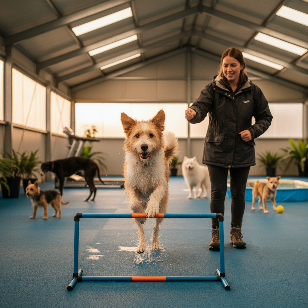
After months of reduced winter exercise, many dogs enter spring with deconditioned muscles, excess body weight, and pent-up energy. When owners suddenly introduce vigorous activities (long hikes, ball chasing, agility courses, or rough play at the dog park), the cranial cruciate ligament bears forces it is not prepared to handle. Veterinary orthopedic literature consistently identifies this pattern of abrupt activity escalation as a primary risk factor for CCL rupture.
Unlike the acute sports injuries seen in human ACL tears, most canine CCL tears involve a degenerative component. The ligament weakens over weeks or months due to subtle fiber damage, obesity, conformational factors, or chronic low-grade inflammation. The dramatic spring outing simply delivers the final mechanical load that completes a partial or full rupture. Breeds commonly reported to be at elevated risk include Labrador Retrievers, Golden Retrievers, Rottweilers, Newfoundlands, and Staffordshire Bull Terriers, though any breed or mixed-breed dog can be affected.
Biomechanics: How the Injury Happens
The cranial cruciate ligament runs diagonally inside the stifle (knee) joint, connecting the femur to the tibia. Its primary role is to prevent the tibia from sliding forward relative to the femur (cranial tibial thrust) and to limit internal rotation and hyperextension of the joint.
The Mechanics of Rupture
During explosive movements such as sudden stops, sharp directional changes, or jumping and landing on uneven terrain, the stifle undergoes simultaneous flexion, internal rotation, and axial loading. In a conditioned joint with a healthy ligament, these forces are distributed across the CCL, the caudal cruciate ligament, the menisci, and the surrounding musculature. In a deconditioned dog with a degenerating CCL, this force combination can exceed the ligament's tensile limit, causing partial or complete rupture.
The Tibial Plateau Angle Factor
Dogs have a naturally sloped tibial plateau (typically around 20 to 30 degrees in most breeds). This slope creates a persistent cranial shear force during weight bearing. The steeper the slope, the greater the demand on the CCL. This anatomical reality explains why CCL disease is far more common in dogs than in cats, and why certain breeds with steep tibial plateau angles are disproportionately affected.
The Contralateral Limb Risk
Veterinary orthopedic studies suggest that a significant proportion of dogs (commonly cited as 40 to 60 percent) that rupture one CCL will go on to injure the opposite leg within one to two years. Limping on the injured leg shifts compensatory load to the contralateral stifle, accelerating degeneration of that ligament. This is an important consideration in rehabilitation planning.
Recognizing a CCL Tear as an Emergency
Many owners delay veterinary evaluation because their dog "still puts some weight on it" or "seems better after resting." This delay can worsen meniscal damage, increase joint inflammation, and complicate surgical outcomes. The following signs warrant urgent veterinary assessment.
Red Flag Signs: Seek Veterinary Care Immediately
- Sudden, acute hind-limb lameness during or immediately after vigorous activity.
- Non-weight-bearing lameness: the dog holds the affected leg up and refuses to place the paw on the ground.
- Audible pop or cry at the moment of injury, followed by reluctance to move.
- Rapid stifle swelling (visible puffiness around the knee joint within the first hour).
- "Toe-touching" stance: the dog barely touches the toe to the ground but will not bear full weight.
- Sitting abnormally: the affected leg is held out to the side rather than tucked under the body (sometimes called a "lazy sit" or "positive sit test").
Signs That Suggest a Partial Tear or Chronic Degeneration
- Intermittent hind-limb lameness that worsens after exercise and improves with rest.
- Stiffness after lying down, particularly noticeable in the morning or after naps.
- Gradual muscle wasting (atrophy) in the affected thigh compared to the opposite side.
- Reluctance to jump onto furniture, climb stairs, or enter vehicles.
Even partial tears are considered veterinary urgencies because they frequently progress to full rupture without intervention. Early diagnosis can significantly improve long-term outcomes.
Immediate First Aid: What to Do in the Next 10 Minutes
A CCL tear is not a life-threatening emergency in the same category as bloat or hemorrhage, but prompt and correct first aid prevents further joint damage and reduces pain.
Step-by-Step Immediate Response
- Stop all activity immediately. Do not allow the dog to continue walking, running, or playing. Carry small dogs; guide large dogs slowly on a short leash.
- Confine the dog to a small, padded area. A crate, a penned-off section of a room, or a car with a flat cargo area works well. The goal is to prevent any jumping, turning, or stair climbing.
- Apply a cold compress if tolerated. Wrap ice or a bag of frozen vegetables in a thin towel and hold it gently against the swollen stifle for 10 to 15 minutes. Never apply ice directly to skin or fur without a barrier.
- Do not administer human pain medications. Ibuprofen, acetaminophen (paracetamol), and naproxen are toxic to dogs. If the dog has a previously prescribed veterinary anti-inflammatory, contact the prescribing veterinarian before giving a dose.
- Call the veterinary clinic. Describe the injury mechanism, the onset of lameness, and the degree of weight bearing. Ask whether the dog should be seen as a same-day urgent case or within 24 hours.
What NOT to Do: Common Dangerous Mistakes
- Do not massage, manipulate, or attempt to "reset" the joint. Forcing flexion or extension on a ruptured CCL can tear the meniscus, dramatically worsening the injury and surgical prognosis.
- Do not apply a splint or bandage to the stifle. Improper bandaging of the hind limb frequently causes pressure sores, circulation compromise, or further instability. Stifle immobilization requires veterinary-grade equipment and expertise.
- Do not allow "leash walking to see if it gets better." Even gentle leash walks generate cranial tibial thrust. Strict crate rest is appropriate until veterinary evaluation.
- Do not give corticosteroids without veterinary direction. While they reduce inflammation, corticosteroids can mask pain, encourage overuse of the damaged joint, and interfere with surgical planning.
- Do not assume improvement equals healing. Dogs with partial CCL tears often appear to improve after 48 to 72 hours of rest, only to rupture completely during the next burst of activity.
Getting to the Emergency Vet Safely
For large dogs, use a blanket or towel as a sling under the abdomen to support hind-limb weight during the walk to the vehicle. Lift from underneath, never by the limbs. In the car, confine the dog to a flat surface; avoid allowing the dog to sit on a slippery back seat where sudden braking could cause further twisting of the stifle. Small dogs can be transported in a secure carrier.
If the injury occurs on a trail or remote area, carry the dog or fashion a makeshift stretcher from a jacket and two sturdy branches. Minimize distance walked on the injured leg. For dogs too large to carry, walk slowly on a very short leash, supporting the hindquarters with a sling. Owners who regularly hike with dogs in spring should consider carrying a commercially available emergency dog sling or harness. For more on safe spring outings, see our guide on training your dog to stay calm around spring wildlife.
What to Tell the Vet on Arrival
Emergency and orthopedic veterinarians rely on accurate owner history to guide diagnostics. Prepare the following information:
- Exactly what the dog was doing when the lameness began (jumping, turning, landing from a height, running on uneven ground).
- Whether there was an audible sound (pop, crack, or yelp) at the time of injury.
- The timeline: how many minutes or hours ago the injury occurred.
- Weight-bearing status: non-weight-bearing, toe-touching, or intermittent lameness.
- Prior lameness history: any previous episodes of hind-limb stiffness, limping, or a diagnosed partial tear.
- Current medications and supplements, especially anti-inflammatories or joint supplements.
- The dog's recent activity level: mention if the dog has been relatively sedentary over winter and recently increased exercise intensity. This context helps the veterinarian assess whether the injury fits a degenerative CCL pattern.
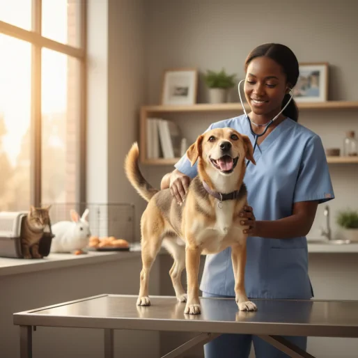
The veterinarian will typically perform an orthopedic examination including the cranial drawer test and the tibial thrust test. Sedation is often necessary for accurate assessment in tense or painful dogs. Radiographs (X-rays) help evaluate joint effusion, arthritis, tibial plateau angle, and rule out fractures. Advanced imaging such as MRI may be recommended in complex or ambiguous cases.
Treatment Options: Surgical vs. Conservative Management
Surgical Repair (Recommended for Most Dogs)
Veterinary orthopedic consensus, supported by organizations such as the American College of Veterinary Surgeons (ACVS), generally favors surgical stabilization for dogs over approximately 10 to 15 kg (22 to 33 lb) with complete CCL rupture. Common procedures include:
- Tibial Plateau Leveling Osteotomy (TPLO): The tibial plateau is cut and rotated to reduce its slope, eliminating the cranial tibial thrust that the CCL normally restrains. This is currently one of the most widely performed and studied CCL surgeries.
- Tibial Tuberosity Advancement (TTA): The tibial tuberosity is advanced forward to change the angle of the patellar tendon force, neutralizing tibial thrust through a different biomechanical approach.
- Lateral Fabellar Suture (Extracapsular Repair): A heavy suture material is placed outside the joint to mimic the CCL's restraint. This technique is more commonly used for smaller dogs or when osteotomy procedures are not available.
Surgical outcomes generally show good to excellent return to function in 85 to 90 percent of cases when followed by appropriate rehabilitation. The choice of technique depends on the dog's size, conformation, activity level, concurrent meniscal injury, and the surgeon's expertise.
Conservative (Non-Surgical) Management
Conservative management may be considered for dogs under approximately 10 to 15 kg, dogs with significant anesthetic risk due to concurrent disease, or cases where owners cannot pursue surgery. It involves:
- Strict rest and activity restriction for 6 to 8 weeks.
- Veterinary-prescribed anti-inflammatory and analgesic medications.
- Weight management (critical: even modest weight loss reduces stifle loading significantly).
- Gradual, controlled physiotherapy.
- Custom orthotic bracing in select cases.
Conservative management typically results in the formation of periarticular fibrosis (scar tissue) that provides partial stabilization, but it does not restore normal joint biomechanics. Progressive osteoarthritis is expected regardless of treatment path, though surgical stabilization generally slows its progression. Dogs managed conservatively often develop chronic, low-grade lameness and are at higher risk for meniscal tears.
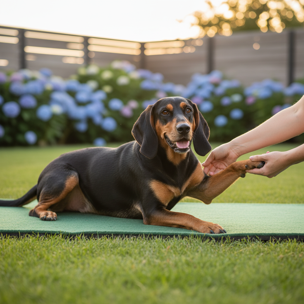
Recovery and Rehabilitation Timeline
Post-surgical rehabilitation is critical and should follow a structured protocol designed or supervised by a veterinary rehabilitation specialist (if available). The following is a general framework; individual plans vary based on the procedure performed, the dog's size, and concurrent injuries.
Weeks 1 to 2: Strict Confinement
- Crate or pen rest with leash walks only for toileting (5 minutes, flat ground).
- Cold compressing the surgical site for 10 to 15 minutes, two to three times daily.
- Passive range-of-motion exercises as directed by the surgeon.
- Incision monitoring for signs of infection (redness, discharge, swelling, heat).
- E-collar (cone) worn at all times to prevent licking.
Weeks 3 to 6: Controlled Leash Walking
- Gradual increase in leash walk duration (from 5 minutes up to 15 to 20 minutes by week 6).
- Introduction of gentle therapeutic exercises: sit-to-stand repetitions, weight shifting, and controlled stepping over low obstacles.
- Hydrotherapy (underwater treadmill) may begin around weeks 3 to 4 if available, providing excellent low-impact muscle strengthening.
- Continued restriction from stairs, jumping, and off-leash activity.
Weeks 7 to 12: Progressive Strengthening
- Leash walks increase to 20 to 30 minutes on varied terrain (gentle inclines, grass, soft ground).
- Balance and proprioception exercises (wobble boards, cavaletti poles).
- Continued hydrotherapy sessions.
- Veterinary recheck with possible follow-up radiographs around weeks 8 to 10.
Weeks 13 to 16 and Beyond: Return to Activity
- Gradual, supervised off-leash activity in controlled environments.
- Slow reintroduction of moderate-intensity exercise (short jogs, easy fetch on flat ground).
- Full return to unrestricted activity is typically not recommended before 16 weeks post-surgery, and some dogs benefit from longer timelines.
- Long-term joint-health strategies: weight management, ongoing low-impact exercise, and veterinary-recommended joint supplements.
For dogs recovering from orthopedic surgery, environmental management is crucial. Senior dogs and those with concurrent arthritis face additional challenges; our article on home physiotherapy for arthritic senior pets covers complementary principles that apply across species. Similarly, owners managing a recovering pet alongside the demands of spring should be aware that senior dogs and cats overheat faster, which can complicate outdoor rehabilitation sessions in warming weather.
Preventing CCL Tears: A Spring Conditioning Plan
The most effective prevention strategy is gradual reconditioning. Veterinary sports medicine professionals recommend the following approach as winter transitions to spring:
- Week 1 to 2: Add 5 to 10 minutes of controlled leash walking per day beyond the winter baseline.
- Week 3 to 4: Introduce gentle incline walking and controlled trotting. Avoid fetch, frisbee, and off-leash running.
- Week 5 to 6: Begin short, supervised off-leash sessions on even ground. Introduce low-intensity play.
- Week 7 onward: Gradually return to full spring activity levels, including longer hikes and moderate-intensity games.
Maintaining a lean body condition year-round is arguably the single most important modifiable risk factor. The Association for Pet Obesity Prevention consistently reports that a majority of dogs in developed countries are overweight or obese, placing chronic excess load on the stifle joints. Dogs adopted from rescues, who may have unknown orthopedic histories, require especially careful conditioning; see our guide on adopting a dog from a breed-specific rescue for additional health-screening considerations.
When to Return to the Emergency Vet After Treatment
Whether managed surgically or conservatively, owners should seek urgent veterinary reassessment if any of the following occur:
- Sudden worsening of lameness after a period of improvement (possible meniscal tear or implant complication).
- Swelling, heat, or discharge at the surgical incision.
- Fever (rectal temperature above 39.5°C or 103.1°F).
- Complete refusal to eat or drink for more than 24 hours post-operatively.
- Acute lameness developing in the opposite hind leg.
- Signs of systemic illness: lethargy, vomiting, pale gums, or rapid breathing.
A Final Word on Urgency
CCL tears are among the most common orthopedic injuries in dogs, and the spring activity surge makes this a peak period for presentations. Early veterinary evaluation, prompt and appropriate first aid, and commitment to a structured rehabilitation program give dogs the best chance of returning to comfortable, active lives. Delaying assessment in the hope that "it will get better on its own" risks meniscal damage, chronic arthritis, and a significantly worse surgical prognosis. When in doubt, treat sudden hind-limb lameness as an urgent veterinary matter.
Frequently Asked Questions
Can a dog's cranial cruciate ligament tear heal on its own without surgery? ↓
How soon after a suspected CCL tear should a dog see a veterinarian? ↓
Why are CCL tears more common in spring than other seasons? ↓
What is the typical recovery time after TPLO surgery for a CCL tear? ↓
If my dog tears one CCL, will the other leg be affected too? ↓

Dr. Ana Reyes
Emergency & Critical Care Veterinarian
Emergency and critical care veterinarian — life-saving first-aid guidance and emergency recognition for pet owners.
Content Disclosure
This article was created using state-of-the-art AI models with human editorial oversight. It is intended for informational and entertainment purposes only and does not constitute veterinary medical advice. Always consult a licensed veterinarian for your pet's specific health needs. Learn more about our process.